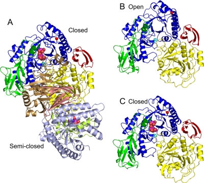
FIGURE 2.
Overall structure of GLNBP. The TIM barrel fold domain
(blue), Ig-like fold domain (green), α/β fold
domain (yellow), and C-terminal domain (red) are shown.
A, dimeric structure of GlcNAc-NO3-EG crystal form.
Subunits in the closed and semiclosed states are shown in a ribbon
model. The semiclosed state subunit is shown with a different color
code (TIM barrel domain in light blue, Ig-like domain in
light green, α/β domain in brown, and C-terminal
domain in pink). The ligand-free form in the open state (subunit A)
(B) and GlcNAc-NO3-EG in the closed state (subunit A)
(C) are shown from the same view as A. Ligands in the active
site (GlcNAc, ethylene glycol, and  )
are shown as a space-filling model. Asp-313 and Trp-233 are shown as
a stick model (cyan), and the Cα atom of Gly-371 is
shown as a red sphere.

