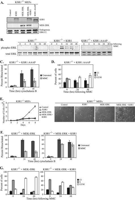
FIGURE 7.
The KSR1-ERK interaction is required for cell cycle reinitiation. A, KSR1–/– or KSR1–/– MEFs expressing KSR1, MEK-ERK, MEK-ERK+KSR1, or KSR1.AAAP were lysed and subjected to Western blotting for the indicated proteins using an anti-KSR1 (top panel) or anti-ERK1/2 antibody (bottom two panels). B, KSR1–/– or KSR1–/– MEFs expressing either WT KSR1 or KSR1.AAAP were assessed for MMC-induced ERK activation. MEFs were treated with 0.5 μg/ml MMC for 2 h, washed with PBS, and incubated for the indicated times in fresh medium. Total and phospho-ERK were assessed by Western blotting. C, KSR1–/– MEFs expressing KSR1.AAAP were treated with 0.5 μg/ml MMC for 2 h (light gray bars) or left untreated (dark gray bars). Following MMC treatment, the cells were incubated with cytochalasin B for the indicated times and analyzed by CBPI assay. The percentage of binucleated cells is indicated. The values are the averages ± standard deviations of three trials. Control KSR1–/– and KSR1–/– MEFs expressing ectopic KSR1 are shown in Fig. 2. D, KSR1–/– MEFs expressing KSR1.AAAP were treated with MMC (0.5 μg/ml) for 6 h and were analyzed by propidium iodide staining and flow cytometry at the indicated times after treatment. The percent of cells in G1 phase (black bars), S phase (gray bars), or G2/M phases of the cell cycle (white bars) are shown. The values are the averages ± standard deviations of three independent trials. Control KSR1–/– and KSR1–/– MEFs expressing ectopic KSR1 are shown in Fig. 3. E, KSR1–/– MEFs (diamonds) or KSR1–/– MEFs expressing either KSR1 (squares), MEK-ERK (triangles), or MEK-ERK+KSR1 (circles) were seeded at 4 × 10⁴ cells/35-mm-diameter dish. Triplicate dishes were assessed for cell number every 48 h on a Beckman Coulter counter. Focus formation assays were performed as described under “Experimental Procedures.” Representative photomicrographs are shown (right panel). F and G, KSR1–/– MEFs expressing MEK-ERK or MEK-ERK+KSR1 were analyzed by CBPI assay (F) or flow cytometry (G) as described above.

