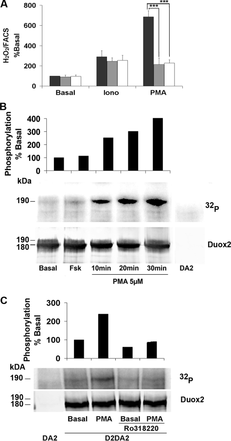
FIGURE 6.
PMA stimulates the activity and phosphorylation of Duox2. A, cells co-transfected with HA-Duox2/DuoxA2 were preincubated 30 min in Krebs-Ringer-Hepes medium containing vehicle (black bars) or PKC inhibitors: 1 μm Ro318220 (gray bars) or 1 μm Gö6976 (open bars) before 2.5 h of stimulation with 1 μm ionomycin (Iono) or 1 nm PMA. H2O2 accumulation was normalized to Duox2 expression at the plasma membrane. The level of H2O2 is represented as a percentage of the value obtained in basal condition without PKC inhibitor (means ± S.D., n = 6). Statistically significant inhibition is indicated. ***, p < 0.001. B, phosphorylation by 32P incorporation measured after 10, 20, or 30 min of treatment with 5 μm PMA. On the top of the Western blot, the relative amount of phosphorylated Duox2 corrected to total Duox2 protein (basal phosphorylation was considered as 100%). C, inhibition of PMA-mediated Duox2 phosphorylation by Ro318220. The cells were preincubated or not with 1 μm Ro318220 before stimulated with 100 nm PMA. Total Duox2 proteins were detected with anti-Duox antibody, and the relative Duox2 phosphorylation corrected to total Duox2 protein is represented at the top of the figure (basal phosphorylation without PKC inhibitor was considered as 100%).

