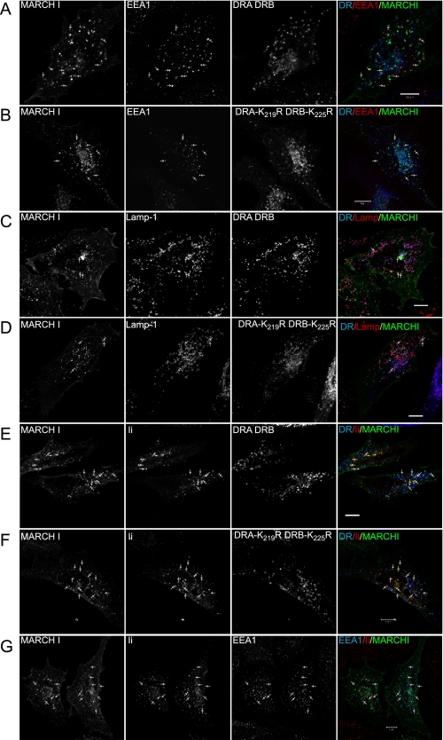
FIGURE 7.
Intracellular distribution of MHC class II and MARCH I. HeLa cells were transiently transfected with wild-type and mutated DRα and DRβ constructs, together with MARCH I, in the presence or absence of Ii. Intracellular distribution was analyzed by confocal microscopy. A and B show colocalization of MARCH I and EEA1 (arrows), in cells transfected with either DRA/DRB (A) or DRA-K219R/DRB-K225R (B). The merged image shows DR in blue, EEA1 in red, and MARCH I in green; co-localized MARCH and EEA1 is yellow. C and D show colocalization of MARCH I and Lamp-1 (arrows), in cells transfected with either DRA/DRB (C) or DRA-K219R/DRB-K225R (D). The merged images show DR in blue, Lamp-1 in red, and MARCH Iin green. Note that the majority of class II is present in Lamp-1-positive compartments (purple), some of which co-localize with MARCH I. E–G show localization of class II in the presence of Ii. The merged image shows DR or EEA1 in blue, Ii in red, and MARCH I in green. E and F show colocalization of Ii and MARCH I (arrows) in cells transfected with DRA/DRB (E) or DRA-K219R/DRB-K225R (F). In both cases, Ii shows good colocalization with MARCH I, whereas DR is mainly in MARCH I-negative vesicles. G shows a high degree of colocalization between Ii and EEA1. Together, this shows that Ii is in MARCH I-positive, EEA1-positive early endosomes. Bar, 10 μm.

