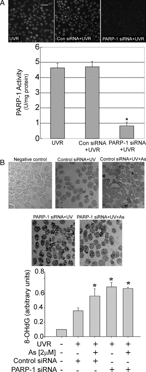
FIGURE 4.
PARP-1 gene silencing increases UVR-induced oxidative DNA damage. A, cells were transfected with control or PARP-1 siRNA as described under “Experimental Procedures.” Upper panel: PARP-1 activity was detected by in situ immunochemical detection of poly (ADP-ribose). Lower panel: PARP-1 activity was measured in cell extracts as described in the legend to Fig. 2C. Values shown represent the mean of three independent experiments ±S.D.; *, p < 0.05 compared with UVR alone. B, cells were transfected with control or PARP-1 siRNA as described under “Experimental Procedures.” Cells were incubated without or with 2 μm arsenite in serum-free medium for 24 h, then exposed to 8 J/cm2 UV radiation and placed on ice. 8-OHdG was measured by immunoperoxidase staining (upper panel) and quantified using image analysis software (lower panel) 120 min after UVR exposure. Each experiment was repeated at least three times. Bars represent the mean ±S.D.; *, p < 0.05 compared with UVR alone. No significant differences were detected between the UVR plus As(III) and UVR plus PARP-1 siRNA groups.

