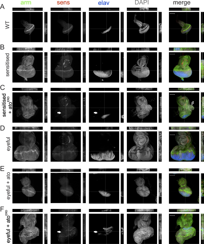
Figure 3. Analysis of Tissue Patterning and Differentiation in Third Instar Eye Discs.
Immunohistochemistry was used for armadillo (arm, green), embryonic lethal, abnormal vision (elav, blue), senseless (sens, red), and diamidinophenylindole (dapi, grey)
(A) Wild-type eye disc.
(B) ey-GAL4, UAS-Dl/+ shows enlarged discs with wild-type patterning.
(C) ey-GAL4, UAS-Dl/UAS-atoERD show disrupted patterning with expansion of the undifferentiated domain (white arrows). Proliferative outgrowth is indicated with an open arrow.
(D) ey-GAL4, UAS-Dl, eyeful/+.
(E) Gain of ato function in the eyeful background leads to restoration of the pattern of differentiation: ey-GAL4, UAS-Dl, eyeful/UAS-ato shows almost normal appearance of all markers.
(F) Loss of ato function in an eyeful background leads to loss of uniform arm staining and a loss of and abnormal pattern of differentiation (elav and sens, white arrows). ey-GAL4, UAS-Dl, eyeful/UAS-atoERD.
Images and orthogonal sections are shown. All images were taken at the depth of the nuclei. Scale bars represent 100 μm.

