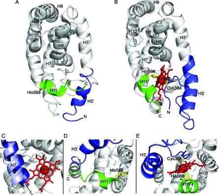
Figure 7. The Structure of REV-ERBβ LBD in Complex with Fe(III) Heme.
(A) Structure of REV-ERBβ LBD without [21] and with (B) heme bound in the ligand-binding pocket.
(C) The position of H7 in the apo- (dark blue [21]) and heme-bound (light blue) states of REV-ERBβ.
Detailed view of heme-binding site of REV-ERBβ LBD in the absence (D) [21] and presence (E) of heme. The core of the protein is in gray, N-terminal part of H3 (H3') is in blue and H11 is in green. The side chains of Cys384 and His568 are shown in yellow and heme is colored red.

