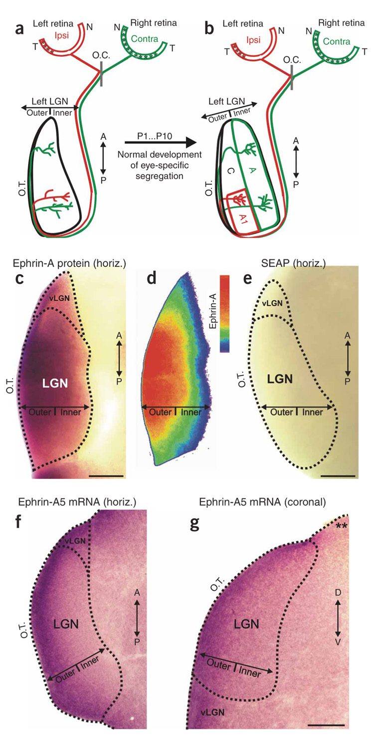
Figure 1. Eye-specific development and ephrin-As in the ferret LGN.
(a,b) Schematic diagram of RGC axon ingrowth and arborization during eye-specific segregation in the P1 and P10 LGN. Horizontal plane is shown and, for simplicity, only the retinal projection to the left LGN is shown. Asterisks (in the retinas) indicate the location of contralateral and ipsilateral RGCs that project to the left LGN. O.C., optic chiasm; T, temporal pole; N, nasal pole; A, contralateral eye layer; A1, ipsilateral eye layer; C, nonprincipal layers; A/P, anterior-posterior axis; O.T., optic tract. (c) Expression of pan-ephrin-A protein in a horizontal section of the P3 ferret LGN. (d) Color-scaled densitometry plot of ephrin-A protein in the P3 LGN. (e) Secreted embryonic alkaline phosphatase (SEAP)-AP shows no staining in the P3 ferret LGN. (f) Outer > inner gradient of ephrin-A5 mRNA expression in horizontal section of the P0 ferret LGN. (g) Outer > inner gradient of ephrin-A5 mRNA seen in a coronal plane section of the P1 ferret LGN. Asterisks indicate an edge of the same tissue section that does not exhibit dioxigenin (DIG) labeling, indicating that the outer > inner gradient in the LGN is not due to edge artifacts. D–V, dorsal-ventral axis; vLGN, ventral lateral geniculate nucleus. Scale bars, 150 µm (c–g).

