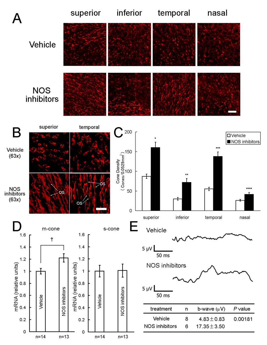
Figure 4. Nitric oxide synthase (NOS) inhibitors promote cone survival in rd1 mice.
Starting at P18, rd1 mice received twice daily intraperitoneal injections of vehicle or vehicle containing a mixture of four NOS inhibitors, L-NNA, L-NAME, L-NMMA, and aminoguanidine. At P35, cone density was measured in 0.0529 mm2 bins 1 mm superior, inferior, temporal, and nasal to the optic nerve, or the retina was used for real time RT-PCR. Confocal images appeared to show a higher density of cone inner segments, particularly superior and temporal to the optic nerve (A). Three-dimensional reconstruction of confocal images showed preservation of cone outer segments (OS) in some NOS inhibitor-treated mice (B, bottom row), whereas all vehicle-treated mice showed small flattened inner segments and no OS (B, top row). Cone density was significantly higher in the NOS inhibitor group (n=9) in all 4 regions of the retina (C, *p<5.0×10⁻⁶; **p<5.0×10⁻⁵; ***p<5.0×10⁻⁸; ****p<0.01 by unpaired Student t-test for difference from corresponding vehicle control (n=17). The mean (±SEM) amount of m-cone or s-cone opsin mRNA per retina was normalized to the P35 vehicle-treated group, which was set to 1.00. The amount of m-cone opsin mRNA, but not s-cone opsin mRNA, was significantly greater in NOS inhibitor-treated mice compared to vehicle-treated mice (D, *p<0.02 by unpaired Student t-test). Photopic electroretinograms done on rd1 mice treated with NOS inhibitors at P25 showed greater b-wave amplitudes than those seen in vehicle-treated mice (E). Scale bars = 50 µm for (A) and 20 µm for (B)

