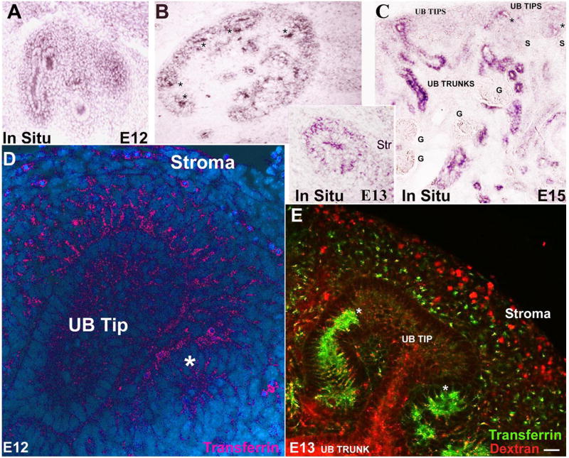
Figure 4.
Localization of the transferrin pathway. (A, B, C) TfR1 was initially localized to UB tips and cap mesenchyme. By E13, renal vesicles also expressed TfR1 (*B, C). TfR1 expression was reduced or absent in glomeruli (“G”), stroma and capsule. (D) Rhodamine- and (E) fluorescein-transferrin endocytosis paralleled TfR1 expression. Stroma and capsule were weakly labeled despite active fluid-phase endocytosis (rhodamine-dextran; E). (*Presumptive distal nephron; Bar= 10 μm).

