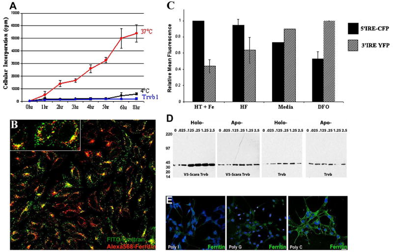
Figure 7.
Scara5 mediates cellular uptake of ferritin-bound iron and represents an endogenous pathway in MSC-1 cells. (A) Capture of 59Fe-ferritin by Scara5+ at 37°C (red line) but not at 4°C (black line) nor by parental Trvb cells (blue line). (B) Lysosomes of Scara5+ Trvb cells were marked with fluorescein-dextran (green). They contained endocytosed Alexa568-ferritin (red). Original magnification 40X. (C) Transfer of iron to the cytoplasm was detected using two IRE-based iron reporters and FACS analysis in MSC-1 cells. The maximal 5′ IRE-CFP signal (designated 100%-black bars) was generated by iron loading with holotransferrin (20 μg/ml) and ferric ammonium citrate (20 μM) (HT+Fe) and the maximal 3′ IRE-YFP signal (designated 100%-stippled bars) was generated by DFO (20 μM). These treatments produced a ~7-fold range in the ratio of 5′/3′ IRE signals. Treatment with holoferritin (HF) increased 5′ IRE-CFP but decreased the 3′ IRE-YFP signal. (D) Holoferritin induces cell growth. Scara5+ clone H responded to holo- but not to apo-ferritin, whereas Trvb cells failed to respond. Cell protein was detected by GAPDH immunoblots. (E) Alexa488-ferritin endocytosis was blocked by Poly I >Poly G >Poly C (50 μM). Similar data were obtained in Scara5+ Trvb and MSC1 cells. Bar=20 μM.

