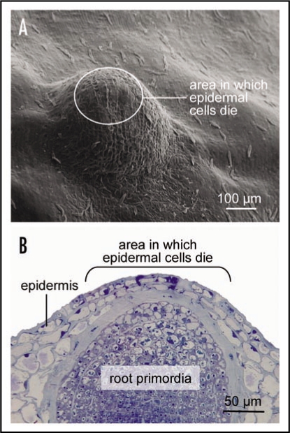
Figure 1.
(A) Scanning electron microscopic view of nodal tissue with an underlying adventitious root primordia. The waxy surface structures differ between epidermal cells above the root primordia which can undergo cell death and other epidermal cells. (B) Light micrograph of a cross section through the node showing a root primodia. Indicated in (A and B) are the approximate areas in which epidermal cells will undergo cell death when triggered by an appropriate signal. Approximately 2,600 genes are differentially expressed in these epidermal cells prior to cell death induction.

