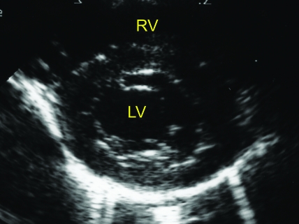
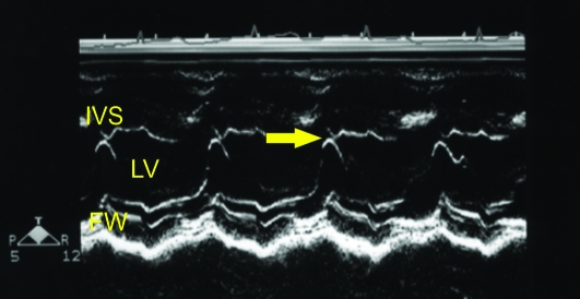
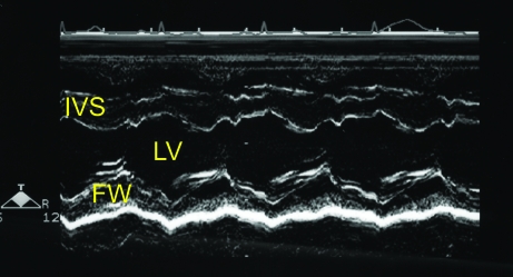
Figure 1.
(A) Right parasternal short axis echocardiogram obtained at the level of the mitral valve annulus. (B) M-mode image obtained from the level depicted in Figure 1A. The E point to septal separation would be obtained from this view (arrow). (C) M-mode image obtained at the ventricular level (slightly ventral to that depicted in Figure 1B). The ventricular dimensions and shortening fraction were obtained from M-mode imaging at this cardiac level. FW, left ventricular free wall. IVS- interventricular septum; LV, left ventricle; RV, right ventricle.


