
Fig. 2.
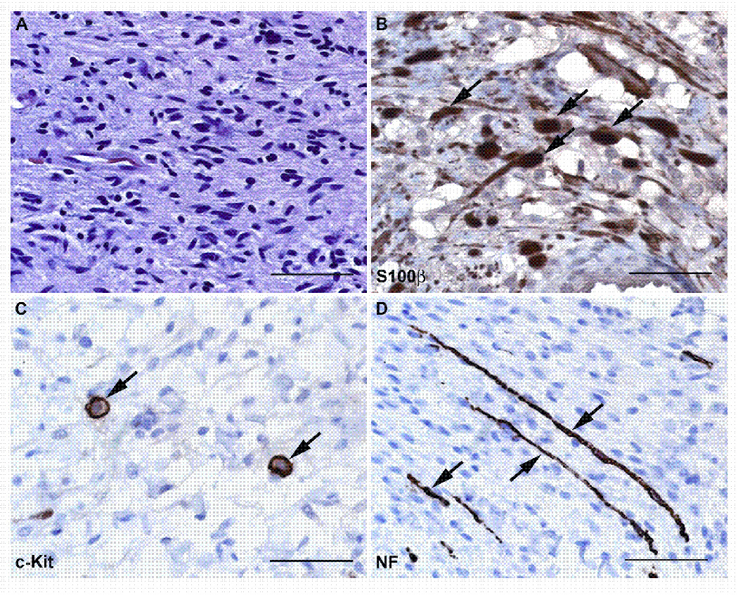
Neurofibromas are composed of a complex mixture of cell types. (A) High power (40x) view of a hematoxylin and eosin stained section from a localized intraneural (nodular) neurofibroma that arose on the 5th lumbar nerve of a 61 year old woman. This neoplasm is composed of a mixture of cell types that can be detected using immunostains for cell-type specific markers, as shown in B–D. (B) Immunoperoxidase stains (brown) for S100β label the Schwann cell-like elements in this neurofibroma. The section has been lightly counterstained with hematoxylin to highlight the nuclei of cells (primarily fibroblasts and perineural-like cells) that do not stain for this antigen. Arrows indicate some of the cells that are intensely S100β immunoreactive. (C) Another section of this tumor immunostained for the mast cell marker c-Kit (CD117). Arrows indicate two cells that show intense membranous immunoreactivity for this membrane tyrosine kinase receptor. (D) The same tumor immunostained for neurofilaments to demonstrate the presence of axons entrapped by the infiltrative growth pattern of this tumor (arrows). Bars in A–D: 50 µm.