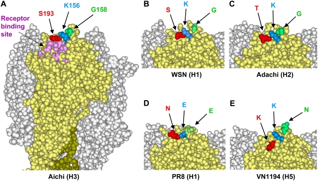
Figure 5. Structure of the MAb S139/1 epitope on the globular head of HA trimer models.
Three-dimensional models of Aichi (H3) (A), PR8 (H1) (D), and VN1194 (H5) (E) HAs were constructed from the coordinates obtained from the Protein Data Bank (PDB codes: 1HGF, 1RVX, and 2IBX, respectively). The structures of WSN (H1) (B) and Adachi (H2) (C) were constructed by homology modeling as described in Materials and Methods. Images were prepared by using DS Visualizer (version 1.7, Accelrys, Inc.). Residue numbering is thoroughly on the basis of the H3 HA sequence.

