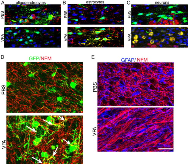
Figure 4.

The lineage choice of transplanted immunoselected A2B5+/GFP+/NG2+ oligodendrocyte progenitors is affected by VPA. Confocal imaging of the medial corpus callosum in P10 rats transplanted with A2B5+/NG2+ progenitors isolated from GFP+ rats is shown. A–C, Note that in PBS-treated controls, the majority of the transplanted GFP+ cells (green) were also CC1+ (A, top, red) but not GFAP+ (B, top, red) or NeuN+ (C, top, red). DAPI (blue) was used as a nuclear stain. In contrast, in VPA-treated recipients, the majority of the transplanted cells were GFAP+ (B, bottom, yellow) or NeuN+ (C, bottom, yellow) but not CC1+ (A, bottom). D, Colabeling of GFP (green) and NFM (red) indicated the presence of double immunoreactivity (arrows) only after VPA treatment. E, Colabeling of GFAP and NFM showed their mutually exclusive expression in the cortex of VPA-treated animals and PBS controls. Scale bars: (in C) A–C, 25 μm; (in E) D, E, 80 μm.
