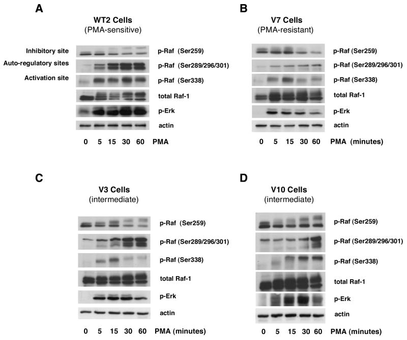
Figure 1. Time course of the acute effects of PMA on Raf phosphorylation in EL4 cell lines.
WT2 (panel A), V7 (panel B), V3 (panel C), and V10 (panel D) cells were treated with or without 100 nM PMA for the indicated times. Immunoblots were performed on whole-cell lysates for phospho-Raf (Ser259, Ser289/296/301, Ser338), c-Raf, phospho-Erk, and actin. All incubations were done in the same experiment; blots for the four cell lines were exposed in parallel.

