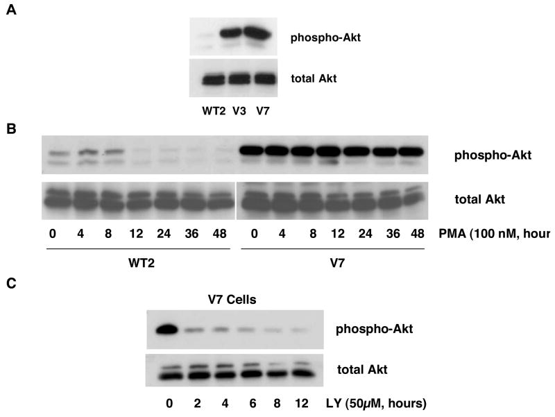
Figure 3. Constitutive Akt phosphorylation in EL4 cell lines.
In panel A, whole-cell extracts from WT2, V3, and V7 cells, equalized for protein, were immunoblotted for phospho-Akt and total Akt. In panel B, WT2 and V7 cells were incubated with 100 nM PMA for indicated times. Whole-cell extracts, equalized for protein, were immunoblotted for phospho-Akt and total Akt. In panel C, V7 cells were incubated with 50 μM LY294002 for indicated times. Whole-cell extracts, equalized for protein, were immunoblotted for phospho-Akt and total Akt.

