
Figure 1.
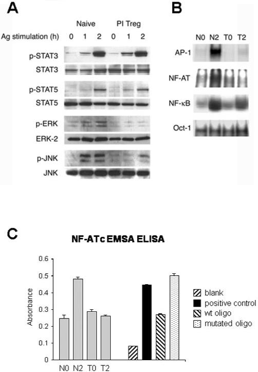
Differential activation of cytokine and T cell receptor signaling pathways in naive and PI-Treg cells. Total CD4+ T cells were isolated from splenocytes of naive or tolerant mice before or 2 h after intranasal stimulation with Ac1-9[4Y]. (A) Activation of STAT and MAP kinases was assessed by immunoblotting with phospho-specific antibodies as indicated. Abundance of STAT3, STAT5, ERK and JNK was quantified by specific antisera for equal loading of protein. Nuclear extracts were analyzed by EMSA (B) using 32P-labeled probes for NFAT, NF-κB, AP-1 and Oct-1 or by ELISA for NFAT (C). The results shown are representative of three separate experiments.