
Figure 4.
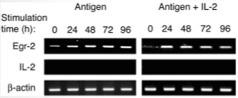
Egr-2 expression in PI-Treg cells following exposure to antigen and antigen + IL-2. Splenocytes from naive and tolerant mice were cultured with Ac1-9[4 K] peptide (10 μg/mL) or peptide + IL-2 (20 U/mL) for 24 h. CD4+ cells were purified from splenocyte cultures using CD4+ microbeads and total RNA was extracted. Transcripts of Egr-2 and IL-2 were semiquantitated by RT-PCR. RT-PCR of β-actin served as loading control. These results were similar in three separate experiments.