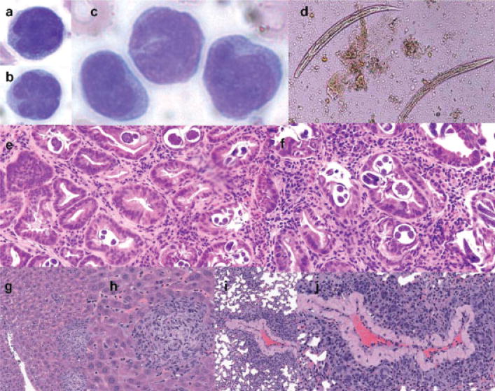
Figure 2.
Pathologic findings. (a–c) Atypical lymphocytes in peripheral blood, and (d) Strongyloides stercoralis in the stool and (e,f) gastric biopsy are shown. (g,h) Liver and (i,j) lung infiltration in NOD.Cg-PrkdcSCIDIL2rgtm1Wjl/SzJ mouse inoculated with PBMCs from the patient. Routine hematoxylin and eosin staining is shown.

