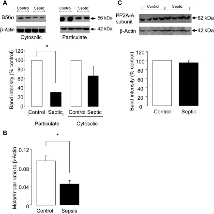
Figure 5.
(A) Representative blot and quantification of the amount of the B56α subunit of protein phosphatase 2A (PP2A) in control and septic hearts separated into particulate and cytosolic fractions (n = 3 hearts/gp, *P < 0.001). Band intensities are normalized to β-actin as a loading control. (B) Amount of mRNA of the B56α subunit present in control and septic hearts determined by RT–PCR (n = 3 hearts/gp, *P < 0.01). (C) Representative blot and quantification of the amount of PP2A-A subunit in septic and control hearts (n = 4 hearts/gp). Band intensities are normalized to β-actin as a loading control.

