Figure 2.
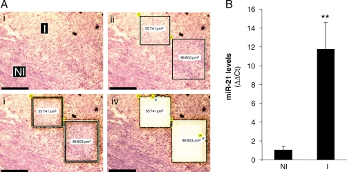
Induction of miR-21 in the infarct region of ischaemia-reperfused murine heart: a laser-capture microdissection based approach. C57BL/6 mice were subjected to ischaemia (30 min) and reperfusion (IR). Heart samples were collected 7 days post-IR. (A) Frozen sections (10 µm) from collected heart samples were stained with haematoxylin QS to histologically define the infarct (I) and control or non-infarct (NI) regions. These regions were subsequently cut and captured using laser-capture microdissection for miRNA expression analysis. (i) Representative images of heart tissue showing infarct (I) and control or non-infarct (NI) areas following laser-capture microdissection compatible staining with haematoxylin QS; (ii) the identified area is marked; and (iii) laser-assisted cutting and separation of the identified area for catapulting; (iv) the section after the marked area has been catapulted; Scale bar = 150 µm. (B) The laser-captured tissue elements from I and NI regions were used for quantification of miR-21 expression using real-time PCR. Data represent mean ± SD (n = 4). **P < 0.01 compared with control (NI) tissue.

