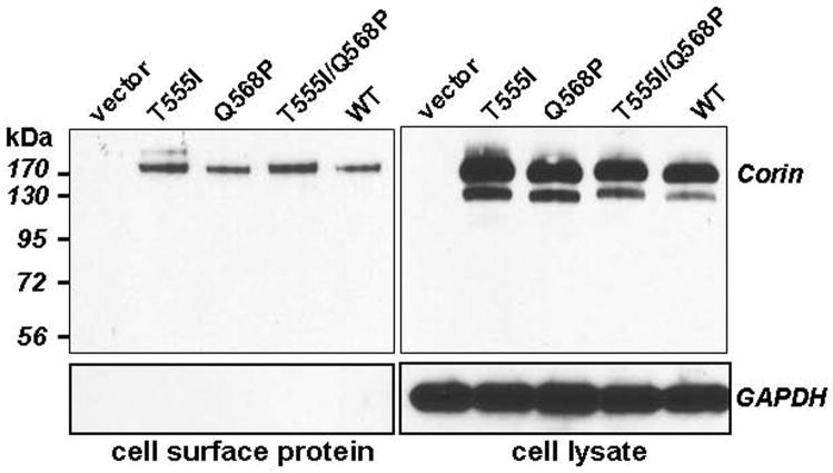
Fig. 4.
Cell surface expression of corin proteins. Cell surface proteins in HEK 293 cells expressing corin proteins or control cells were biotinylated and analyzed by Streptavidin precipitation followed by Western blotting using an anti-V5 antibody (top left). As a control, total corin proteins in cell lysates were verified using the same antibody (top right). As another control for cell surface protein labeling, the same Western blots were analyzed by an antibody against GAPDH (lower panels). The results shown were representative of four independent experiments.

