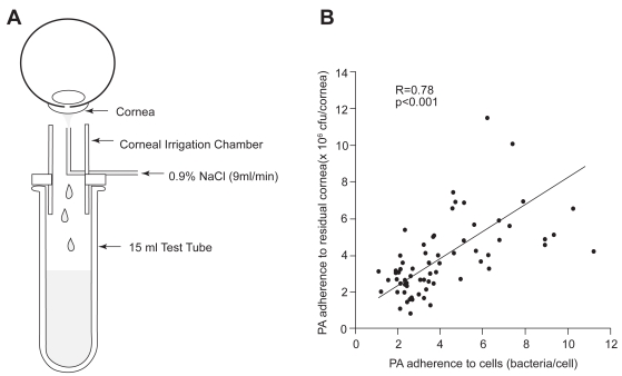
Figure 4.
(A) Schematic of the noninvasive ocular irrigation device. Warmed saline is gently applied to the eye and collected in a test tube directly below. Cells are then incubated in an invasive strain of PA for 30 minutes, vacuum filtered onto polycarbonate filters, and stained with acridine orange for visualization using an epifluorescent microscope. (B) Correlation of PA adherence between exfoliated corneal epithelial cells and the residual corneal surface (R = 0.78, P < 0.001). Copyright © 1997. Images adapted with permission from Ren DH, Petroll WM, Jester JV, et al. 1997. Adherence of Pseudomonas aeruginosa to shed rabbit corneal epithelial cells after overnight wear of contact lenses. CLAO J, 23:63–8.

