Figure 1.
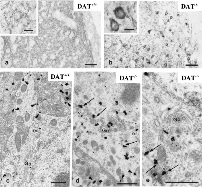
Immunohistochemical detection of D1R at the light and electron microscopic level in DAT+/+ (a and c) and DAT−/− mice (b, d, and e). In wild-type mice (DAT+/+) (a and c), D1R is located in the neuropile and at the plasma membrane of cell bodies (a, Inset; and c, arrowheads). Stars point to dendrite profiles. Ultrastructural localization of D1R in c shows very few intracytoplasmic immunoparticles associated with the Golgi apparatus (Go) and the endoplasmic reticulum (er). In DAT−/− mice (b, d, and e), D1R is located primarily in the cytoplasm of cell bodies (b, Inset). The receptor is associated with the Golgi apparatus (Go), the endoplasmic reticulum (arrows and er), and some vesicles (v). A few immunoparticles are detected along the plasma membrane (d, arrowheads). In e, part of D1R immunoreactivity associated with endoplasmic reticulum (arrows) is in the pericentriolar area (asterisk). [Scale bar = 50 μm (a and b), 10 μm (a and b, Insets), and 1 μm (c, d, and e)].

