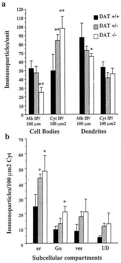
Figure 2.
Quantitative analysis of the subcellular distribution of D1R at the electron microscopic level. (a) Measure of D1R immunoreactivity at the plasma membrane and in the cytoplasm: The number of immunoparticles +/− SEM was counted in relation to the plasma membrane length (Mb IP/100 μm) and to the cytoplasmic surface (Cyt IP/100 μm2) in cell bodies and dendrites. DAT−/− neurons show a sharp and significant decrease of D1R at the plasma membrane of cell bodies and dendrites as compared with DAT+/+ neurons. They also display sharp increase of intracytoplasmic immunoparticles in the cell bodies. DAT+/− mice display significant increase of D1R in the cytoplasm without significant reduction at the plasma membrane. (b) Measure of D1R immunoreactivity in the cytoplasmic organelles in the cell bodies. In DAT+/+ cell bodies, the largest part of cytoplasmic D1R is detected in association with the endoplasmic reticulum (er) and the Golgi apparatus (Go). Other particles are present in vesicles (ves) or in an unidentified cytoplasmic compartment (UD). Immunoparticles density is dramatically increased in the endoplasmic reticulum and in the Golgi apparatus in DAT−/− mice and only in the endoplasmic reticulum in DAT+/− mice. **, P ≤ 0.01; *, P < 0.05.

