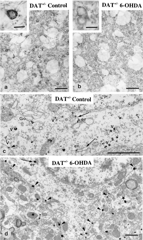
Figure 3.
Immunohistochemical detection of D1R at the light and electron microscopic level in heterozygous mice unilaterally treated with 6–0HDA. In the control side (a and c), striatal neurons display D1R mostly located in the cytoplasm, associated with the Golgi apparatus (Go), the endoplasmic reticulum (er), and vesicles (arrows). In the 6-OHDA-injected side (b and d), D1R appears homogeneously distributed in the neuropile and largely redistributed at the plasma membrane of cell bodies (b, Inset; and d, arrowheads) with a limited number of immunoparticles in the cytoplasm. Star points to a dendritic profile. (Magnification bar: a and b = 50 μm; a and b, Insets = 10 μm; c and d = 1 μm).

