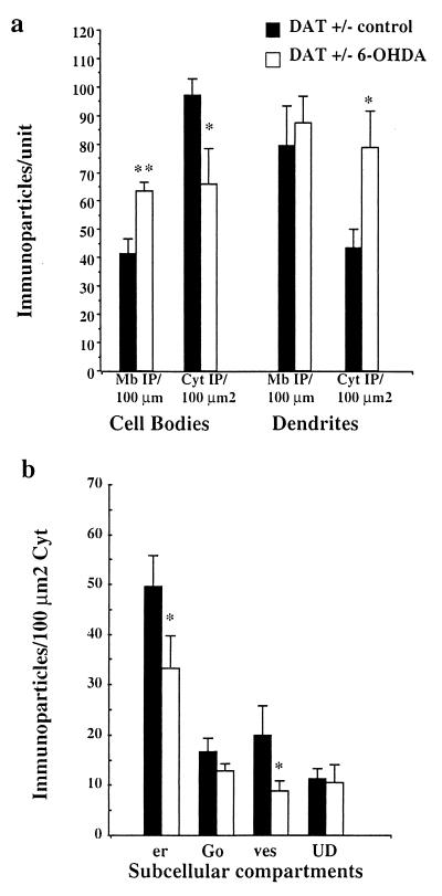
Figure 5.
Quantitative analysis of the subcellular distribution of D1R in DAT+/− mice after 6-OHDA injection. (a) Measure of D1R immunoreactivity at the plasma membrane and in the cytoplasm: The number of immunoparticles +/− SEM was counted in relation to the plasma membrane length (Mb IP/100 μm) and to the cytoplasmic surface (Cyt IP/100 μm2) in cell bodies and dendrites. Cell bodies in 6-OHDA-injected side display decreased cytoplasmic D1R and increased D1R at the plasma membrane. Dendrites display unchanged membrane-bound receptor but increased cytoplasmic D1R. (b) Measure of D1R immunoreactivity in the cytoplasmic organelles in the cell bodies: the endoplasmic reticulum (er) and the vesicles (ves) display a significant decrease of D1R in 6-OHDA-injected side as compared with the control side. **, P ≤ 0.001; *, P ≤ 0.05.

