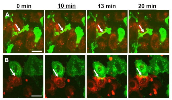
Figure 2.

Lipid mixing of HIV-1 Env-expressing HeLa cells and CD4-X4-expressing NIH3T3 cells in the absence (A) or presence (B) of gp41e. For lipid mixing, HIV-1 Env-expressing (effector) and CD4-X4-expressing (target) NIH3T3 cells were labeled with the 20 μM DiO (green) and DiI (red) probes, respectively. DiI-labeled target cells were co-cultured with DiO-labeled effector cells at 37°C, and lipid dye mixing was monitored microscopically as described in the Materials and Methods. The effector cells co-cultured with target cells exhibited lipid dye mixing at about 13 minutes upon addition of gp41e (B) or not (A), and the intensity continued to increase up to 20 minutes (arrow). Scale bar is 10 μm.
