Figure 3.

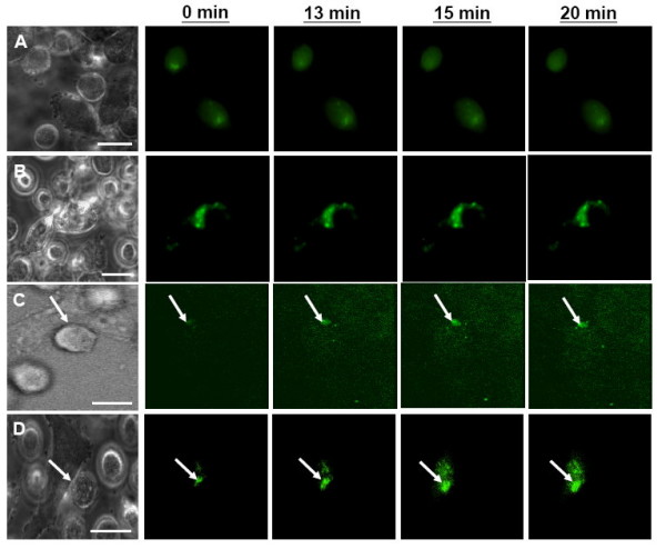
Recruitment of HIV-1 Env-EGFP (green) on the effector HeLa cells upon engagement of CD4-X4-expressing target NIH3T3 cells in the presence of gp41e added at different time points of 0 (A), 10 (B), 13 (C) and 20 minutes (D). Fluorescent images were taken from the same fields of Env-EGFP-expressing HeLa cells (the first bright field) upon addition of target cells. Only images at 0, 13, 15, and 20 minute time points are shown. The movies version is available in Additional Movie 3. After contacting with adherent CD4-X4 cells, HIV-1 Env-EGFP recruitment was observed only at the time points of gp41e added at 13 (C, arrow) and 20 minutes (D, arrow). Scale bar is 10 μm.
