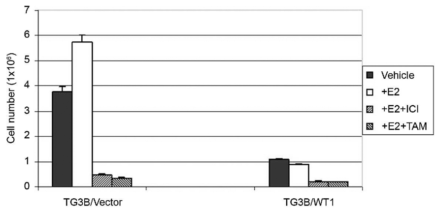
Figure 2. WT1 expression suppresses estrogen-stimulated cell growth in MCF10AT3B cells.
TG3B/vector and TG3B/WT1 cells maintained in phenol red-free DMEM/F12 medium containing 2.5% charcoal/dextran-stripped fetal calf serum were seeded at a density of 1x×103 cells per well in a 6-well dish and treated with 1 nM 17β-estradiol (+E2), or ethanol vehicle as controls (vehicle). After incubation with E2 for 12 days, cells were trypsinized and counted. Antiestrogen, 1 µM of tamoxifen (TAM) or ICI 182, 780 (ICI) was included in control experiments, respectively. Three wells were used for each experiment, which were repeated three times. SD for each point is also shown.

