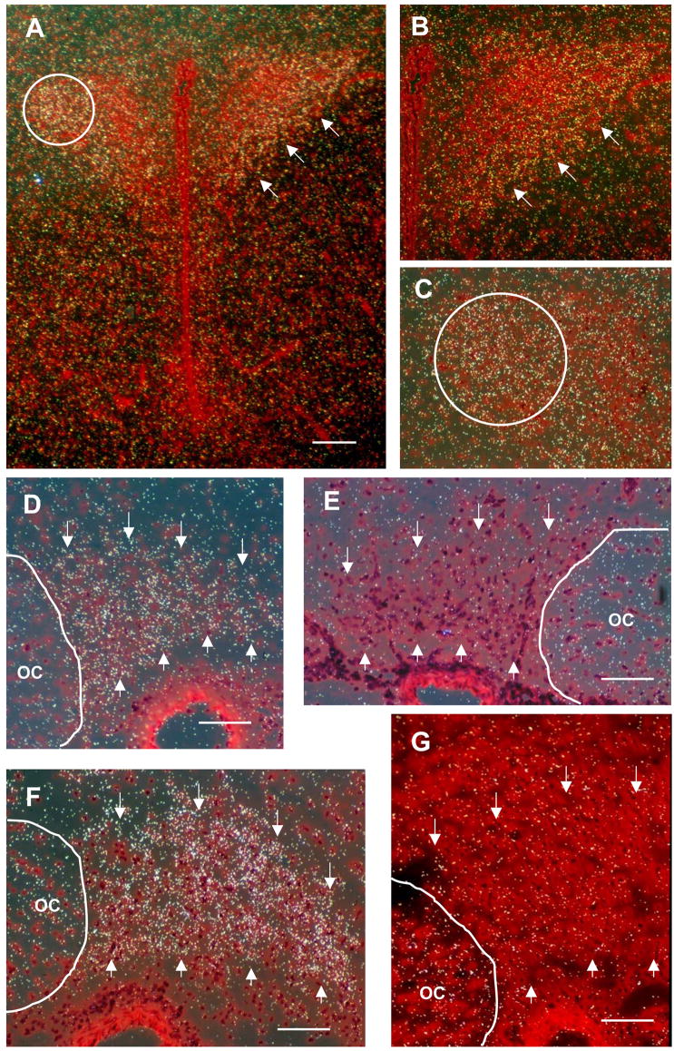
Figure 2.
In situ hybridization revealed that CNTFRα message was expressed in magnocellular divisions of the PVN and in magnocellular neurons in the SON. (A) This low magnification image of the intact (non-lesion control) PVN shows dense grain distributions located predominantly in the lateral magnocellular division (PVL, circle) and the medial magnocellular division (PVM, arrows) of the PVN. Higher magnification images from the same section shown in (A) further illustrate the distribution of grains in the PVL (B) and PVM regions (C, circle). Note the relatively low grain densities in juxtaposed parvocellular regions of the PVN and the surrounding neuropil of the hypothalamus. (D) The SON of intact non-lesion controls, outlined with arrows, also showed significantly higher grain densities than the surrounding neuropil indicative of high levels of endogenous receptor expression. (E) Sense strand control section from the same animal as in D showing grain densities comparable to background levels. (F) Grain densities within the sprouting SON contralateral to the lesion are significantly higher at 7 days post-lesion ( p< 0.05) than in intact control SON. (G) Sense strand control from the same 7 day post-lesion animal shown in F. OC = optic chiasm. Magnification bars: A = 200 μM, D–G = 100 μM.

