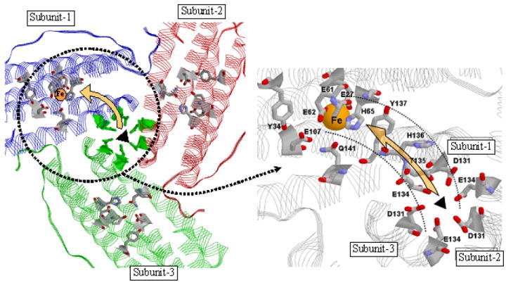
Figure 10.

View from inside of the protein showing the suggested pathway for iron movement from the 3-fold channel to the ferroxidase center (left panel). Expanded view (right panel). The triads of 3-fold channel residues Asp131 and Glu134 are indicated. For clarity the three His118 and three Cys130 residues at the outer opening of the 3-fold channel are not shown. The ferroxidase center ligands His65, Glu27, Glu61, Glu107 and Glu62 and the residues Thr135, His136 and Tyr137 suggested to be involved in shuttling Fe2+ from the channels to the ferroxidase center are shown on the right. Residues Gln141 and Tyr34 involved in H-bonding in Figure 2 are also shown. The single Fe atom at the A-site (His-65 site) of the ferroxidase center is depicted in yellow.
