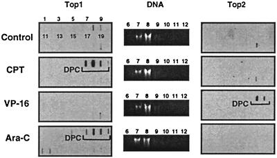
Figure 3.
Top1 cleavage complexes in human leukemia CEM cells treated with ara-C. Approximately 106 cells were treated with 10 μM ara-C for 15 h or with 1 μM CPT or 100 μM VP-16 for 1 h at 37°C. Cells were then lysed with 1% sarkosyl, and subjected to the ICE bioassay (see text for details). Cesium chloride fractions are indicated by numbers 1–20. (A) Top1 immunoblotting. (B) DNA staining of fractions 6–12 with ethidium bromide after electrophoresis. (C) Top2 immunoblotting. Presence of covalent topoisomerase cleavage complexes is indicated by the brackets.

