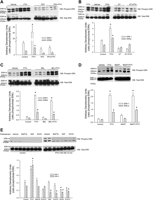
Fig. 4.
Effect of Src kinase, PLC, and PLA2 on PTH-stimulated ERK phosphorylation. OK proximal tubule cells were treated for 15 min with 100 nM PTH in the presence or absence of the Src kinase inhibitor PP2 (100 nM; A), the PLC inhibitor ET (30 μM; B), the iPLA2 inhibitor BEL (1 μM; D), the cPLA2 and iPLA2 inhibitor MAFP (1 μM; D), or the calcium inhibitors BAPTA-AM (20 μM), SKF-96365 (25 μM), or EGTA (1 mM) (E). Cells were lysed, and the lysate proteins were separated by 10% SDS-PAGE, transferred, and immunoblotted for phospho-ERK1/2. Nitrocellulose membranes were stripped and reprobed for total ERK using ERK2 antibodies. A representative blot from 3 different experiments is shown. Bar diagrams show arbitrary densitometry units as the ratio of phosphorylated to total ERK as means ± SE (n = 3). *P < 0.05 by ANOVA followed by Bonferroni analysis.

