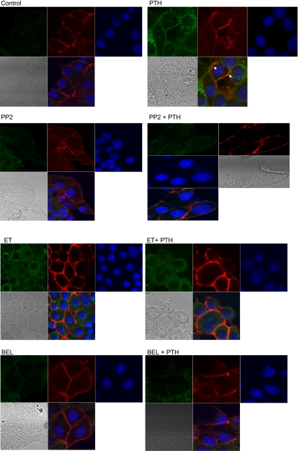
Fig. 5.
Effect of Src kinase, PLC, and PLA2 on PTH-stimulated PKCα translocation. OK cells were plated on 8-well confocal tissue culture plates. Cells were treated for 15 min with 100 nM PTH in the presence or absence of the Src kinase inhibitor PP2 (100 nM), the PLC inhibitor ET (30 μM), or the PLA2 inhibitor BEL (1 μM). Cells were fixed, permeabilized, stained with anti-Na+-K+-ATPase α1-subunit (red) and anti-phospho-PKCα (green), and visualized using a Zeiss confocal microscope. Representative confocal pictures are shown (n = 3). Arrows show colocalization of Na+-K+-ATPase α1-subunit with phosphorylated PKCα.

