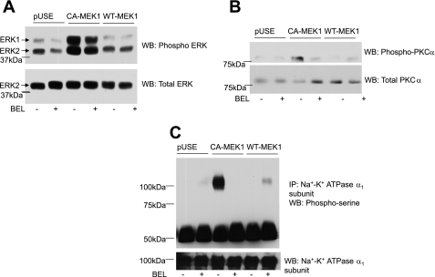
Fig. 6.
Effect of MEK-1 transfection and PLA2 on PKCα and Na+-K+-ATPase α1-subunit phosphorylation. OK cells were transiently transfected with empty vector (pUSE) or constitutively active (CA-MEK1) or wild-type MEK-1 (WT-MEK1) as described in methods. Cells were lysed 24 h after transfection and subjected to 10% SDS-PAGE (A–C) for Western blot analysis for phospho-ERK1/2 (A) and phospho-PKCα (B). Samples from the same lysates were analyzed for Na+-K+-ATPase α1-subunit phosphorylation by immunoprecipitation as described above (C). A representative blot from 2 independent experiments is shown. Western blots are shown for phospho-ERK1/2, phospho-PKCα, and phosphoserine. Nitrocellulose membranes were stripped and reprobed for total ERK, PKCα, and Na+-K+-ATPase α1-subunit (n = 2).

