
Figure 2. cPLA2 in the tumor microenvironment is essential for lung cancer progression.
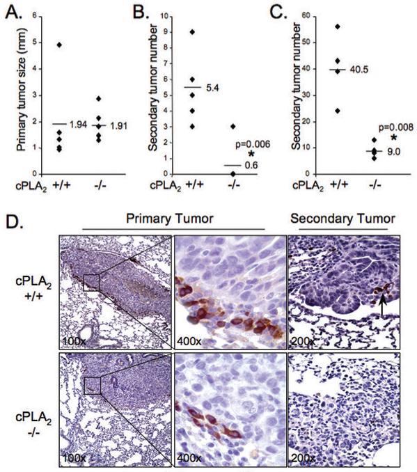
105 CMT167 cells were injected into 5 WT or 5 cPLA2-KO mice as above. Lungs were harvested 4 wks after injection. (A). Primary tumor size (diameter) was measured by caliper. Each point represents a single animal. (B). Numbers of large secondary tumors on left and right lungs were determined. Each point represents a single animal. Four cPLA2 KO mice had no secondary tumors. (C) 105 LLC cells were injected into 4 WT or 4 cPLA2-KO mice. Lungs were harvested as described above, and primary and secondary tumors evaluated. (D). Lung sections from WT (top panels) and KO (bottom panels) mice injected with CMT/167 cells were immunohistochemically stained for F4/80, a macrophage marker (brown reaction color). Arrow = F4/80(+) cells in secondary tumors growing in WT mice. Insets in left panels shown at higher magnification in middle panels.