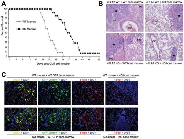
Figure 3. Increased survival and alterations in TAMs surrounding tumors grown in WT and cPLA2-KO mice transplanted with cPLA2 KO bone marrow.

(A). Wild type mice (N=60) were lethally irradiated and transplanted with bone marrow from either UBI-EGFP/B6 WT or cPLA2-KO mice. Five weeks after transplant, mice were inoculated with CMT167 cells. Kaplan-Meier survival curve of WT mice of the indicated bone marrow genotype is shown. (B). H&E stain for tumors and surrounding stroma 4 weeks after injection with CMT167 cells in either WT or cPLA2-KO mice transplanted with the indicated bone marrow. * = primary tumor; AW = airway; PA = pulmonary artery. (C). Immunofluorescence for GFP and F4/80 in bone marrow-transplanted mice. Lung sections from the indicated mice transplanted with either WT or cPLA2-KO bone marrow were examined for accumulation of macrophages (F4/80+) and bone marrow-derived cells (GFP+). Mice receiving UBI-EGFP/B6-derived WT bone marrow were examined for GFP (green) and stained for F4/80 (red). Double positive cells were detected surrounding the primary tumor (Left-most panels, top and bottom). Mice receiving cPLA2-KO bone marrow were stained for F4/80 to detect macrophages (Right-most panels, top and bottom).
