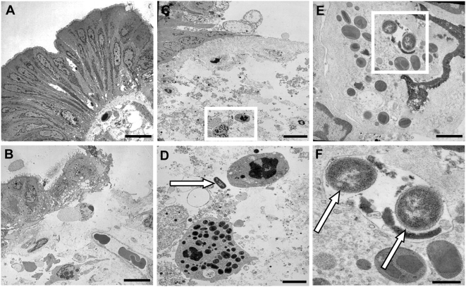
Figure 1. Transmission electronic microscopy of human colonic mucosa infected or not with S. flexneri (M90T).
(A) Control tissue shows normal epithelial lining with regular basolateral folds and absence of bacteria in the lamina propria. (B) Tissue infected with M90T shows regions of partially detached and vacuolated epithelial cells or regions with complete detachement of epithelial lining. (C) Tissue infected with M90T showing regions of detached epithelial cells and presence of bacteria within the lamina propria adjacent to granulocytes (white square) (D). Magnified view of the selected area from C showing bacteria (arrow) close to granulocytes. (E) Granulocyte containing an intracellular vacuole filled with bacteria (white square). (F) Magnified view of the selected area from E showing two intracellular bacteria (arrows). Bar: 10 µm (A, B, C), 2 µm (D), 1 µm (E), 0.5 µm (F).

