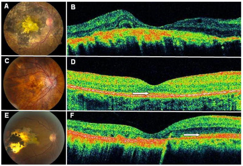
Figure 4. Status of Photoreceptor Layer in Relation to Atrophic Changes at the Level of the Retinal Pigment Epithelium and Choroid.

The right eye of patient 12 (A) with had severe RPE/choroid disease with absence of the overlying photoreceptors (B). The right eye of patient 13 (C) with had milder RPE disease, and the overlying photoreceptors were partially intact (D). The right eye of patient 2 (E) revealed a distinct boundary between intact photoreceptors overlying healthy appearing RPE/choroid and absent photoreceptors overlying atrophic RPE/choroid (F). White arrows denote the photoreceptor layer. OCT mages represent horizontal line scans through center of fovea.
