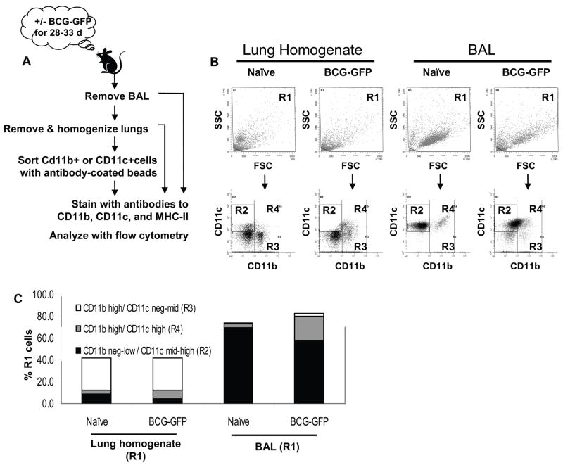
Figure 2. Definition of lung APC subsets by differing expression of CD11b and CD11c.
C57BL/6J mice were aerosol-infected with BCG-GFP for 28–33 d. Lungs were lavaged to obtain BAL cells, perfused with PBS and homogenized. Lung homogenate was analyzed directly or used to prepare CD11b or CD11c affinity-sorted cells. Cells were stained for CD11b, CD11c and MHC-II. Fig. 2 shows flow cytometry analysis of lung homogenate, but all other figures show analysis of CD11b or CD11c affinity-sorted cells, which provide lower autofluorescence background for studies to detect BCG-GFP+ events. A. Sample preparation schematic. B. Dot plots showing forward scatter (FSC) vs. side scatter (SSC) and CD11b vs. CD11c for naïve and BCG-GFP-infected lung homogenate and BAL. The R1 gate was drawn on FSC vs. SSC plots to exclude only subcellular debris in the lower left corner. Cells from R1 were analyzed for CD11b and CD11c to define 3 major APC populations (R2, R3 and R4 gates, see Table 1). C. Mean percentage of R1 cells represented by each APC subsets in lung homogenate and BAL from naïve and BCG-GFP-infected mice. Data was drawn from 3–4 experiments with 2–5 mice per group.

