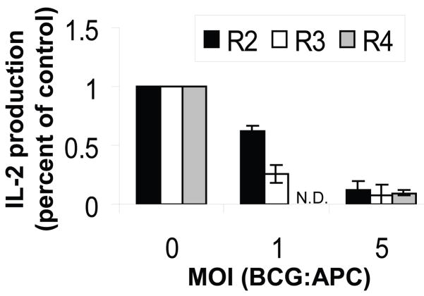
Figure 7. MHC-II antigen processing and presentation by purified lung APCs is inhibited by in vitro infection with BCG-GFP.
Alveolar macrophages (R2), lung macrophages (R3) and lung DCs (R4) were purified by FACS from naïve BALB/c mice and infected with BCG-GFP for 48 h with IFN-gamma (4 ng/ml) present during the final 24 h. Soluble OVA (1 mg/ml) was then added for 24 h, and the APCs were fixed and processed for detection of OVA presentation to DOBW T hybridoma cells. IL-2 production was determined by ELISA. Data represent pooled results of two independent experiments expressed as percent of control IL-2 production observed with each uninfected APC type. Data points represent the mean of four results (two for DCs). DC data were obtained only at MOI of 0 and 5 due to limiting cell numbers. N.D. = not determined.

