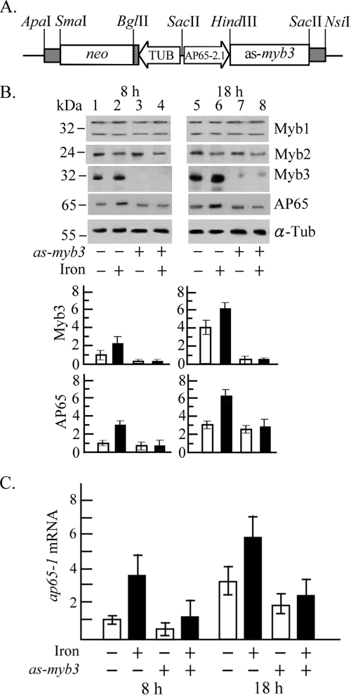
FIG. 4.
Myb3 knockdown in T. vaginalis. (A) In pAP65-2.1as-myb3, the ap65-2 proximal promoter (AP65-2.1) drives an antisense myb3 gene, and the β-tubulin (TUB) proximal promoter drives the neo gene, a selective marker. (B and C) T. vaginalis T1 (lanes 1, 2, 5, and 6) and transfected cells (lanes 3, 4, 7, and 8) were cultured under iron-depleted (open bars; lanes 1, 3, 5, and 7) and iron-replete (closed bars; lanes 2, 4, 6, and 8) conditions for 8 h (lanes 1 to 4) and 18 h (lanes 5 to 8). (B) Total cell lysates were analyzed by Western blotting using the antibodies against Myb1, Myb2, Myb3, AP65, and α-tubulin (α-Tub). Relative levels of Myb3 or AP 65 versus α-Tub are depicted in the bottom panel. (C) Relative levels of ap65-1 versus β-tubulin mRNA in 10 μg RNA from myb3 knockdown (as-myb3) or nontransfected T. vaginalis with iron depletion (open bars) or iron repletion (closed bars) for 8 or 18 h were analyzed by RT-qPCR. The results depicted in bar graphs are the averages ± the standard error from three separate experiments.

