FIG. 7.
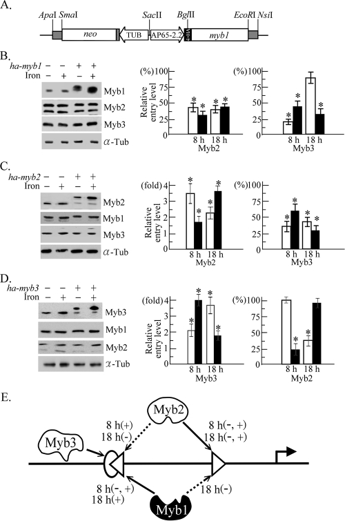
Competitive promoter entries by multiple Myb proteins in T. vaginalis. (A) In pAP65-2.2ha-myb1, the ap65-2.2 promoter (AP65-2.2) drives a myb1 gene with four copies of the HA tag, and the β-tubulin (TUB) promoter drives the neo gene, a selective marker. (B to D) Total lysates (left panel) or cells in a 50-ml culture (middle and right) from T. vaginalis T1 and transfected cells overexpressing HA-Myb1 (ha-myb1) (B), HA-Myb2 (ha-myb2) (C), and HA-Myb3 (ha-myb3) (D) under iron-depleted (open symbols) and iron-replete (closed symbols) conditions for 8 and 18 h were evaluated by Western blotting using the anti-Myb1, anti-Myb2, anti-Myb3, and anti-α-tubulin (α-Tub) antibodies (left) and by ChIP (middle and right). Relative levels of DNA pulled down by the anti-Myb2 and anti-Myb3 antibodies versus input DNA were determined by qPCR. The original levels of Myb2 and Myb3 promoter entry in wild-type cells is considered 1 or 100% to illustrate the relative Myb promoter entry levels in transfected cells. The results are the average ± the standard error from three separate experiments. The data in each set of experiments were analyzed by analysis of variance. A significant difference (P < 0.05) in promoter entry under the same situation in transfected versus wild-type cells is indicated by an asterisk. (E) Competitive promoter entries by Myb1, Myb2, and Myb3 into the MRE-1 (oval), MRE-2r (left-pointing triangle), and MRE-2f (right-pointing triangle) sites in region I of the ap65-1 promoter are depicted. The arrow with a solid line indicates a primary entry site for Myb1, Myb2, and Myb3, and the arrow with a broken line indicates the secondary entry site for a defined Myb protein under iron repletion (+) or iron depletion (−) for 8 or 18 h as indicated. The transcription activator and repressor are depicted by open and closed symbols, respectively.

