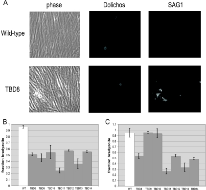
FIG. 1.
The Tbd− mutants show a range of phenotypes. (A) An immunofluorescence assay was used to score the ability of each mutant to differentiate in vitro. After 4 days of bradyzoite induction, the cultures were fixed and parasites were labeled with fluorescein Dolichos, which stains the cyst wall, and anti-SAG1, for the major tachyzoite surface antigen. Parasite cysts are identified by Dolichos staining and an absent or reduced SAG1 signal, while tachyzoite vacuoles lack Dolichos staining and express abundant SAG1. (B) The indicated Tbd− mutants were compared to the WT with respect to their ability to switch from the tachyzoite to bradyzoite form when grown for 96 h in differentiation medium (low CO2, low FCS, high pH). The fraction of each culture that switched to a bradyzoite pattern of gene expression was assessed by binding of the Dolichos bifloros lectin (specific for the cyst wall surrounding bradyzoites) or antibodies specific for the tachyzoite-specific antigen SAG1. Each bar represents two biological replicates with three technical replicates each, and error bars denote the standard deviation. (C) The same Tbd− mutants as in panel B were analyzed for differentiation in response to 900 nM atovaquone. Details are as described for panel B.

