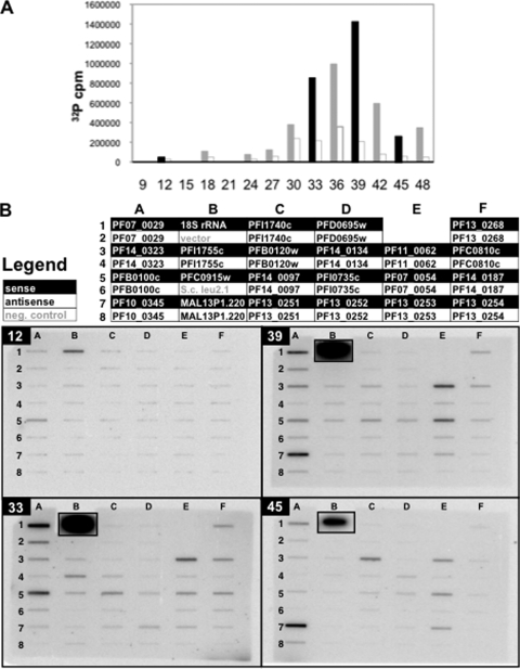
FIG. 3.
Nuclear run-on gene-specific hybridization. (A) Total-incorporation nuclear run-on of time course D is depicted as in Fig. 1, with time points which were assayed by gene-specific nuclear run-on (T12, T33, T39, and T45) shown in black. x axes represent hours postreinvasion. (B) Single-stranded DNA probes were slot blotted onto membranes, which were hybridized with run-on-labeled [32P]RNA from time points T12, T33, T39, and T45 and imaged by exposure of a phosphorimager screen. The legend describes which RNA strand was detected. rRNA slots were cut from their filters and exposed separately to avoid interference between their signals and those of adjacent slots but are shown inset in their original positions.

