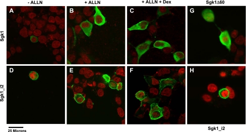
Fig. 5.
Subcellular localization of Sgk1, Sgk1_i2, and Sgk1Δ60. A–F: HEK293 cells grown on chamber slides were transfected with Sgk1-FLAG or Sgk1_i2-FLAG and Sgk1 isoforms were detected by fluorescence confocal microscopy using an anti-FLAG monoclonal antibody (green). Nuclei are stained with To-Pro3 (red). In the absence of ALLN, Sgk1 expression is relatively weak (A) and in the presence of ALLN, there is increased abundance of Sgk1 (B) and the additional treatment with dex does not alter Sgk1 intensity or localization of Sgk1 (C). Neither ALLN nor dex has any apparent effect on Sgk1_i2 (D, E, and F). Overall, Sgk1 is distributed homogeneously throughout the cytoplasm, whereas Sgk1_i2 is predominantly located on the plasma membrane with some distribution seen in cytoplasm. G and H: comparison of Sgk1 where the 1st 60 amino acids are deleted (Sgk1Δ60) with Sgk1_i2 shows that Sgk1Δ60 (G) has a predominantly cytosolic distribution with some nuclear staining also seen, whereas Sgk_i2 (H) is localized to the cell membrane.

