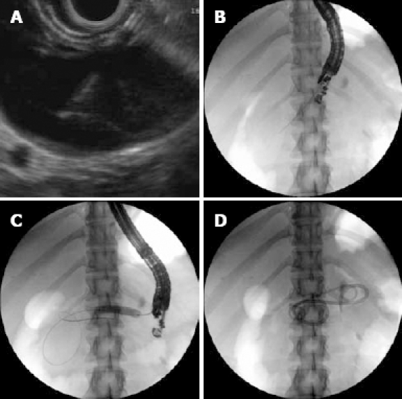
Figure 1.

EUS and fluoroscopic image. A: EUS image of pseudocyst with FNA needle; B: Fluoroscopy image of pseudocyst with FNA needle; C: Fluoroscopy image of balloon dilating the cyst gastrostomy tract; D: Fluoroscopic image of two double pigtail stents draining the pseudocyst cavity.
